Quantitative analyses of the 3D nuclear landscape recorded with super-resolved fluorescence microscopy
Zusammenfassung
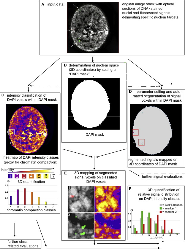
Recent advancements of super-resolved fluorescence microscopy have revolutionized microscopic studies of cells, including the exceedingly complex structural organization of cell nuclei in space and time. In this paper we describe and discuss tools for (semi-) automated, quantitative 3D analyses of the spatial nuclear organization. These tools allow the quantitative assessment of highly resolved different chromatin compaction levels in individual cell nuclei, which reflect functionally different regions or sub-compartments of the 3D nuclear landscape, and measurements of absolute distances between sites of different chromatin compaction. In addition, these tools allow 3D mapping of specific DNA/RNA sequences and nuclear proteins relative to the 3D chromatin compaction maps and comparisons of multiple cell nuclei. The tools are available in the free and open source R packages nucim and bioimagetools. We discuss the use of masks for the segmentation of nuclei and the use of DNA stains, such as DAPI, as a proxy for local differences in chromatin compaction. We further discuss the limitations of 3D maps of the nuclear landscape as well as problems of the biological interpretation of such data.